

About Our Urology Department
Advanced Urological Care at SRM Prime Hospital, Ramapuram
At SRM Prime Hospital, Ramapuram, our Urology Department delivers advanced, evidence-based, and minimally invasive care for the full spectrum of urological conditions — from kidney stones and prostate disorders to urinary tract infections, bladder disease, urological cancers, and complex reconstructive urology. As a leading urology hospital in Chennai, we bring together exceptional surgical expertise, cutting-edge technology, and a deeply patient-centred philosophy to transform outcomes across western Chennai and beyond.
Our department is led by Prof. Dr. R. Manikandan — Clinical Lead and Senior Consultant in Urology with over 20 years of specialist experience, an M.Ch. in Urology from JIPMER Pondicherry, and internationally recognised fellowships in robotic uro-oncology (USA), renal transplantation (Spain), and reconstructive urology (Germany). With over 400 kidney transplants and more than 500 robotic surgeries to his credit, Prof. Dr. Manikandan brings a level of surgical depth and complexity management that is rare outside major tertiary centres.
From your first consultation — whether for a kidney stone, urinary symptoms, prostate concern, or a complex oncological diagnosis — SRM Prime Hospital's Urology Department provides a complete, personalised pathway of care. Our commitment is to restore urinary health, preserve organ function, and return patients to their normal lives with the least possible disruption.
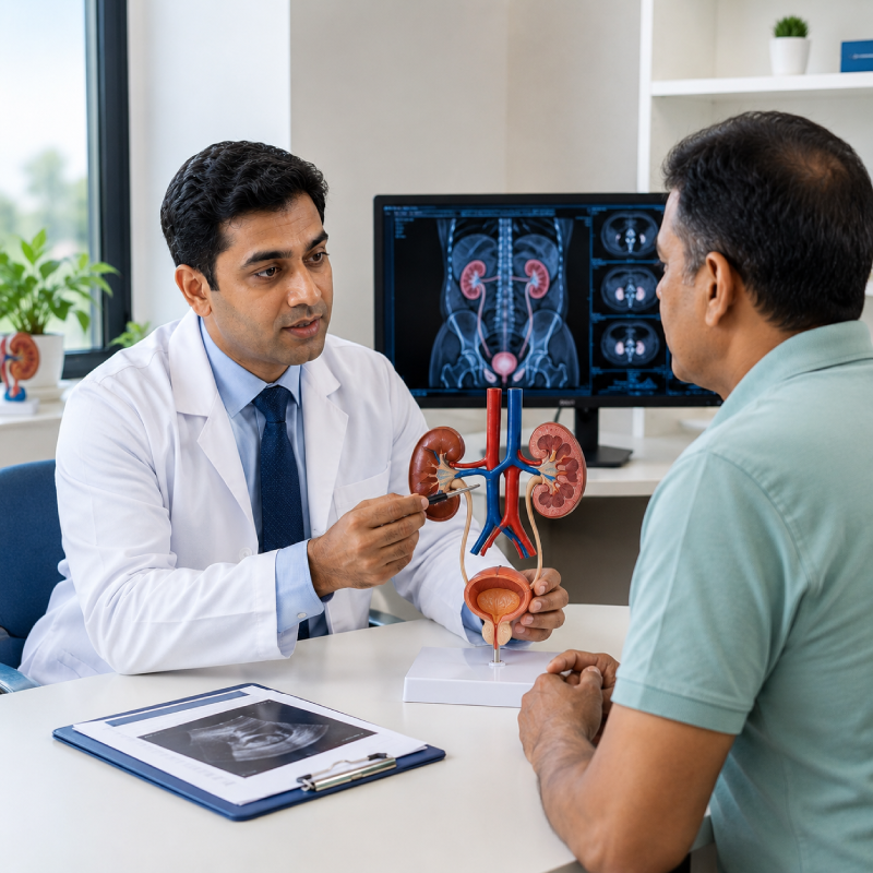
Why Choose Us
Why Choose SRM Prime Hospital for Urology Care
Experience world-class, patient-first care backed by advanced technology, expert specialists and a commitment to better outcomes.

Clinical Lead with internationally recognised fellowships in robotic uro-oncology (USA), renal transplantation (Spain), and reconstructive urology (Germany) — bringing tertiary-centre expertise to western Chennai.

Comprehensive stone disease management — URS, RIRS, PCNL, and laser lithotripsy (MOSES 2.0 Holmium) — for stones of all sizes and locations, from kidney to ureter to bladder.

Robotic-assisted radical prostatectomy, radical nephrectomy, radical cystectomy, and urinary diversion — delivering precision cancer surgery with reduced blood loss, shorter hospital stay, and faster recovery.

One of Tamil Nadu's most experienced urological surgeons — with a volume and complexity of practice across kidney transplant, robotic surgery, and reconstructive urology that rivals leading academic centres.

TURP, laser TURP (HoLEP), MOSES 2.0 laser enucleation for BPH — offering durable, bleeding-minimised prostate treatment with faster catheter removal and return to normal voiding.
Round-the-clock emergency management for acute urinary retention, urosepsis, haematuria, renal colic, urological trauma, and testicular torsion — with dedicated urology emergency protocols.
300+
Beds of Advanced Care
30+
Medical Specialities
7
Modular Operation Theatres
75+
Specialised ICU Beds
Meet Our Urology Team
Our Urology Department is led by one of Tamil Nadu's most accomplished urological surgeons — a clinician, academic, and innovator whose career spans over two decades of complex urological practice at national and international levels.

Prof. Dr. Manikandan R
Clinical Lead & Senior Consultant – Urology
20+ years of experience

Tamil • English

10:00 - 16:00
•
Mon–Sat

Dr. Suresh G
Consultant – Urology
9+ years of experience
English • Tamil
10:00 - 16:00
•
Mon–Sat
Key Highlights
Specialised Urology Services at SRM Prime Hospital
Complete medical and surgical urological care
Flexible ureteroscopy with MOSES 2.0 / Holmium laser for kidney stones up to 2 cm. The flexible scope navigates the ureter and renal collecting system, accessing stones in all calyceal locations without any incision. Stones are fragmented to dust and cleared. Day-care procedure for eligible patients. Preferred for stones not amenable to ESWL and as an alternative to PCNL for selected cases.
Conditions Explained
Conditions We Commonly Treat
Kidney stones (renal calculi or urolithiasis) are crystalline deposits that form within the renal collecting system when urinary solutes — calcium, oxalate, uric acid, struvite, or cystine — precipitate and aggregate. Urolithiasis affects approximately 12% of the Indian population over a lifetime, with recurrence rates of 50% at 5 years without preventive intervention. Tamil Nadu, with its hot climate and dietary patterns high in oxalate-rich foods, has particularly high stone prevalence. Stones range from a few millimetres (passing spontaneously) to staghorn calculi occupying the entire renal pelvis and calyces.
Severe, colicky flank pain (renal colic) — often described as the worst pain a patient has experienced; pain radiates from the flank to the groin, scrotum, or labia as the stone descends the ureter; nausea and vomiting; haematuria — frank (visible) or microscopic blood in urine; urinary urgency and frequency (lower ureteric stones); inability to find a comfortable position (unlike peritonitis, patients are restless); fever and rigors if infection supervenes (infected obstruction — urological emergency); and asymptomatic incidental stones on imaging.
Emergency: urine dipstick (haematuria), urine culture (exclude infection), serum creatinine (renal function), renal ultrasound, and non-contrast CT KUB (gold standard) — identifies all stone types, size, location, and degree of obstruction. Metabolic workup for recurrent formers: 24-hour urine chemistry and stone composition analysis. Treatment by stone size and location: <5 mm — conservative (hydration, alpha-blocker for ureteric stones); 5–10 mm ureteric — URS + laser lithotripsy; <2 cm renal — RIRS (MOSES 2.0 flexible ureteroscopy); >2 cm renal — PCNL. Infected obstructed kidney: emergency drainage (nephrostomy or ureteric stent) followed by definitive stone clearance after infection resolution.
Diagnostic Services
Advanced Urology Diagnostics
Accurate urological diagnosis depends on the right investigation at the right time. SRM Prime Hospital provides a comprehensive urological diagnostic platform — all coordinated by Prof. Dr. Manikandan to ensure clinical efficiency and diagnostic precision.

Gold standard for kidney stone detection — identifies all stone types (including uric acid stones invisible on plain X-ray), stone size, location, HU density, and degree of ureteric obstruction. Also used for trauma, renal mass characterisation, and pre-surgical planning.
Evaluation of haematuria — assessment of renal parenchyma, upper tract urothelium (TCC), and bladder. Renal mass characterisation (enhancement pattern). Pre-surgical planning for complex stone disease and upper tract reconstruction.

Multiparametric MRI with PI-RADS reporting — T2W, DWI (ADC map), and DCE sequences for prostate cancer detection, localisation, and staging (capsular invasion, seminal vesicle involvement). Used for targeted prostate biopsy planning and active surveillance monitoring.

First-line imaging — hydronephrosis, renal cysts (Bosniak classification), prostate volume and morphology, residual urine (PVR), scrotal Doppler (varicocele, torsion, epididymo-orchitis), penile Doppler with vasoactive injection for ED evaluation.

Non-invasive assessment of voiding function — maximum flow rate (Qmax), voiding time, voided volume, and flow curve pattern. Plateau pattern = stricture; prolonged low flow = BPH. PVR: >150 ml = significant retention. Essential before BPH surgery and stricture repair.

Full urodynamic assessment for voiding dysfunction — filling cystometry (bladder sensation, compliance, detrusor overactivity); pressure-flow study (Bladder Outlet Obstruction Index, detrusor contractility); sphincter EMG; and video-urodynamics for neurogenic bladder and complex incontinence.

Definitive imaging for urethral strictures — location, length, calibre, and multiplicity. MCU provides proximal extent and simultaneous bladder assessment. Essential pre-operative evaluation for urethroplasty planning.

DTPA diuretic renogram — split renal function and drainage assessment for UPJ obstruction and post-pyeloplasty follow-up. DMSA scan — cortical scar assessment for VUR-related renal damage and differential renal function before nephrectomy.

PSA (total, free:total ratio, PSA density, PSA velocity) for prostate cancer screening and monitoring. AFP, beta-HCG, LDH for testicular cancer staging. Urine NMP-22 and cytology for bladder cancer surveillance. 24-hour urine stone chemistry for metabolic evaluation.
Direct endoscopic visualisation of urethra and bladder under local anaesthesia — evaluation of haematuria, recurrent UTI, LUTS, bladder cancer surveillance, stent assessment. Blue-light (photodynamic) cystoscopy for enhanced tumour detection in bladder cancer follow-up.
Urological Emergencies
24/7 Care for Urological Emergencies


Frequently Asked Questions
The best treatment for a kidney stone depends on its size, location, composition, and whether it is causing obstruction or infection. Small ureteric stones (<5 mm) often pass spontaneously with hydration and an alpha-blocker (tamsulosin). Larger ureteric stones are treated with ureteroscopy (URS) and laser lithotripsy. Kidney stones up to 2 cm are managed with RIRS (flexible ureteroscopy with MOSES 2.0 laser) — a scarless, day-care procedure. Stones >2 cm or staghorn calculi require PCNL. At SRM Prime Hospital, we have Tamil Nadu's MOSES 2.0 laser platform — delivering the most advanced stone fragmentation available, with superior stone clearance rates and minimal patient discomfort.
No. Mild to moderate BPH symptoms are initially managed medically — with alpha-blockers (tamsulosin) to relax the prostate smooth muscle and improve flow, and 5-alpha reductase inhibitors (finasteride) to reduce prostate volume. Combination therapy and mirabegron for overactive bladder symptoms are also used. Surgery is recommended when symptoms are severe (IPSS >20), when medical treatment fails, or when complications develop — such as acute urinary retention, recurrent UTI, bladder stones, or hydronephrosis. At SRM Prime Hospital, we offer MOSES 2.0 HoLEP (the most advanced laser prostate surgery) and bipolar TURP — both highly effective with durable results.
BPH (benign prostatic hyperplasia) is a non-cancerous enlargement of the prostate that causes urinary symptoms — it does not spread or become cancer. Prostate cancer is a malignant tumour of the prostate that can spread to lymph nodes and bone if not treated. The two conditions can coexist. A PSA blood test and digital rectal examination are the initial screening tools — but a definitive diagnosis of prostate cancer requires a prostate biopsy. Elevated PSA can occur in both BPH and prostate cancer — the decision to biopsy is guided by PSA level, PSA density, PSA velocity, and multiparametric MRI (mpMRI) findings.
RIRS (Retrograde Intrarenal Surgery) uses a flexible ureteroscope passed through the natural urinary passage (no skin incision) to reach kidney stones directly and fragment them with a laser (MOSES 2.0 or Holmium) to dust. It is ideal for kidney stones up to 2 cm, hard stones not amenable to ESWL, and patients with anatomical variations. PCNL (Percutaneous Nephrolithotomy) uses a small skin incision to access the kidney directly — it is preferred for large stones (>2 cm), staghorn calculi, and when RIRS would require multiple sessions. The choice depends on stone burden, location, hardness, and patient factors — your urologist will recommend the optimal approach.
Any visible blood in the urine (frank haematuria) should be investigated urgently, even if it occurs only once and resolves spontaneously. It is a cardinal symptom of bladder cancer, kidney tumours, and kidney stones. Significant microscopic haematuria (≥3 red blood cells per high-power field on microscopy) in adults over 40 also warrants evaluation. Investigations include urine cytology, flexible cystoscopy, CT urogram, and PSA. Do not assume painless haematuria is due to a UTI — haematuria in the absence of UTI symptoms should always prompt specialist urological assessment.
Yes. SRM Prime Hospital has a fully functional renal transplant programme led by Prof. Dr. R. Manikandan, who has personally performed over 400 kidney transplants. We perform living-related and living-unrelated donor transplants, as well as cadaveric (deceased donor) transplants through the Tamil Nadu TRANSTAN network. Our programme covers the complete transplant pathway — from pre-transplant evaluation (cross-match, HLA typing, donor and recipient workup) through laparoscopic donor nephrectomy, recipient transplantation, and long-term immunosuppression management.
Yes. Prof. Dr. R. Manikandan is a fellowship-trained robotic uro-oncologist (USA) with over 500 robotic surgeries to his credit. Robotic-assisted procedures available include radical prostatectomy (for prostate cancer), partial nephrectomy (kidney tumour — nephron-sparing), radical nephrectomy (large renal tumours), radical cystectomy with urinary diversion (bladder cancer), and pyeloplasty (UPJ obstruction). Robotic surgery offers superior visualisation, precision, and dexterity in confined pelvic and retroperitoneal spaces — translating into reduced blood loss, nerve sparing, and faster recovery.
MOSES 2.0 (Modulated Optimised Energy System) is the most advanced Holmium laser platform for urological surgery — available at SRM Prime Hospital as Tamil Nadu's first. It delivers laser energy in a modulated, pulse-modulated waveform that significantly reduces retropulsion during stone fragmentation (meaning stones don't bounce away from the laser fibre) and minimises bleeding during HoLEP prostate surgery. For stone patients, MOSES 2.0 means superior stone fragmentation to fine dust (reduced re-treatment rate), shorter operating time, and better stone-free rates at a single session. For BPH patients, it enables safe HoLEP on large prostates (>100 ml) with minimal bleeding, early catheter removal (24–48 hours), and rapid return of normal voiding.
Blog & Articles
Latest Updates & Health Insights
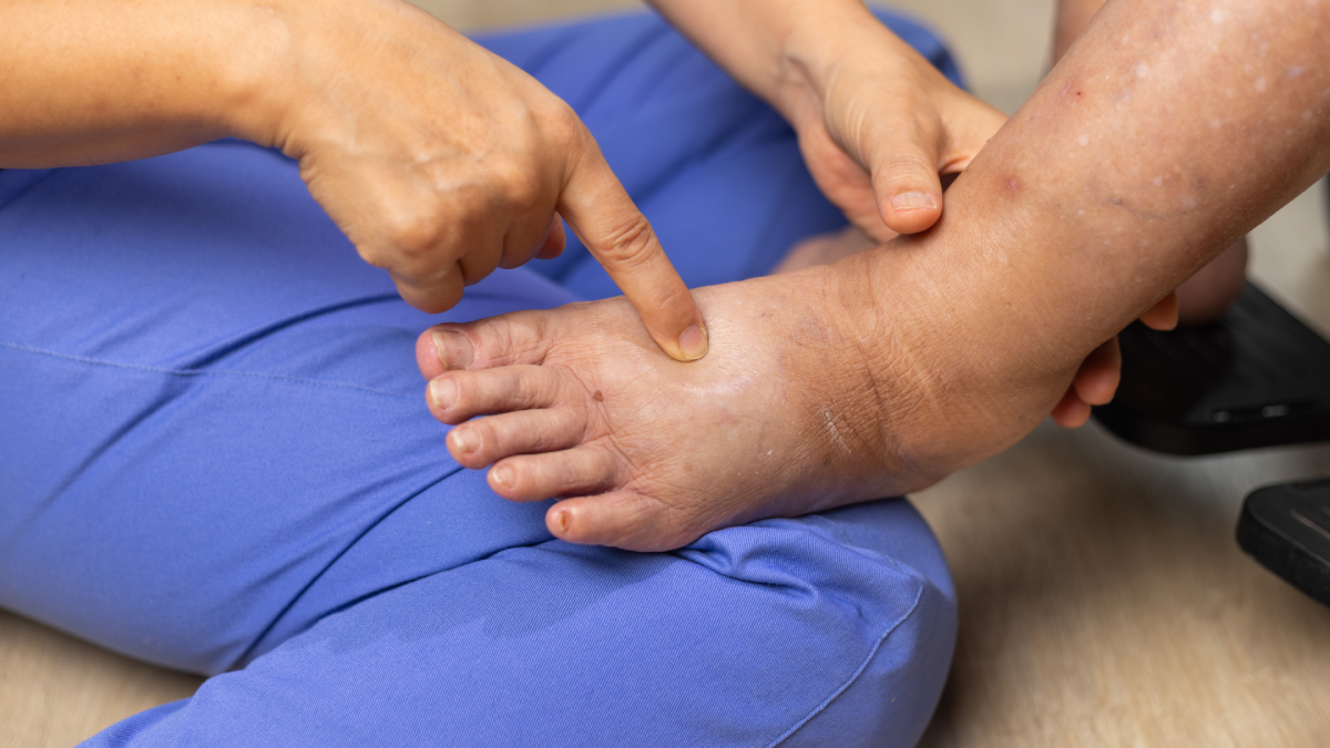
Recognising Diabetic Foot Problems Before They Get Worse
Learn about diabetic foot symptoms, early warning signs, daily checks and when changes like wounds, swelling or colour changes need medical care.
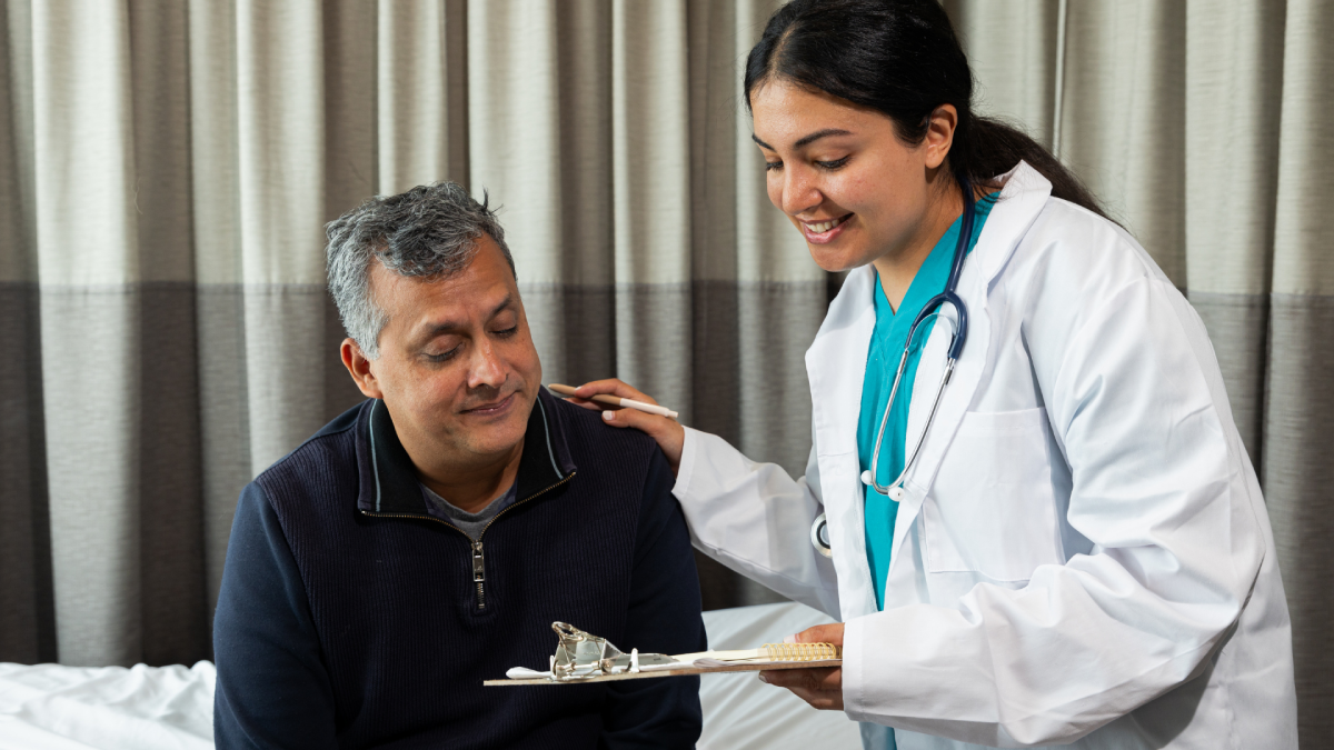
How to Choose the Right Specialist at SRM Prime Hospital
Learn how to choose the right SRM Prime Hospital specialist doctors based on symptoms, diagnosis, medical history and the specialist care you may need.
SRM PRIME Zone: Free Healthcare Benefits for Families Within 5 km of Ramapuram
SRM PRIME Zone: Get your free SRM PRIME Card if you live within 5 km of Ramapuram. 2 free specialist consults, 20% off lab tests, free ambulance and more.

Vomiting: Causes in Adults and Children and When It Becomes Serious
Learn the common vomiting causes in adults and children, warning signs, dehydration risks, and when vomiting needs prompt medical attention and evaluation.

How to Book an Appointment at SRM Prime Hospital
Learn how to book an appointment at SRM Prime Hospital, choose the right specialist, prepare medical records, and plan your consultation with confidence.
Acidity Symptoms: When Everyday Heartburn May Be Signalling More Than Indigestion
Learn about acidity symptoms, common triggers, warning signs and when recurring heartburn may need medical assessment for reflux or GORD.
Request a Callback
Can’t Find What You’re Looking For?